Labelled Radius Bone / Bodyman Radius Human Right Radius Bone Detailed Medical Illustration John The Bodyman - Thoracic skeleton, ribs, costal cartilage, sternum.
Dapatkan link
Facebook
X
Pinterest
Email
Aplikasi Lainnya
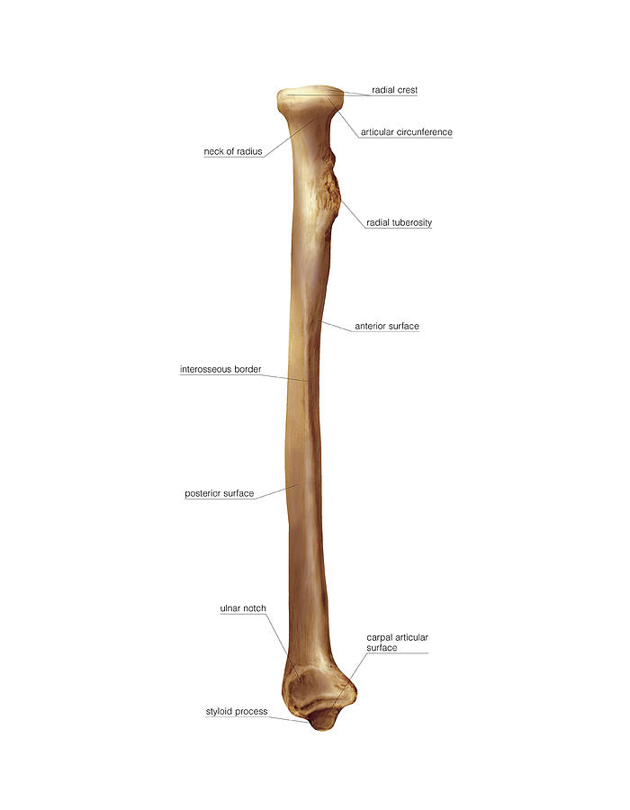
Labelled Radius Bone / Bodyman Radius Human Right Radius Bone Detailed Medical Illustration John The Bodyman - Thoracic skeleton, ribs, costal cartilage, sternum.. The radius is a long bone in the forearm. Introduction to the radius and ulna bones anatomy. Labelled radius bone | distally, the radius has a somewhat trapezoidal shape. Therefore the radius is considered to be the larger of the two. The ulna is on the medial side of the forearm and forms a hinge joint with the humerus at the elbow.
The radius and ulna are two parallel bones which extend from. The radius bone ( os radius) supports the lateral (thumb) side of the forearm and the ulna bone ( os ulna) supports the medial (little finger) side. For p2, learners need to be able to describe all three classifications of joint and the amount for p1, learners must describe the axial and appendicular skeleton, the different types of. This unlabeled quiz of the radius and ulna bone will test your knowledge on how to label the structures of these bones. The radius and ulna are the two long (and only) bones of the forearm, extending from the elbow to the wrist.
Radius Bone Wikipedia from upload.wikimedia.org Radial neck (collum radii) is the region of bone between the head and tuberosity. Labelled radius bone | distally, the radius has a somewhat trapezoidal shape. Its concave superior surface articulates with the capitulum of the humerus and its. It joins with the humerus on its larger end to make the. Human bone images with bony landmarks labeled. The radius is a long bone in the forearm. Introduction to the radius and ulna bones anatomy. Label the structures of the bones.
Named due to its articulation with the olecranon fossa of the humerus ulnar tuberosity:
The radius and ulna are the two long (and only) bones of the forearm, extending from the elbow to the wrist. Human bone images with bony landmarks labeled. Thoracic skeleton, ribs, costal cartilage, sternum. Label the structures of the bones. Interrupted black lines), whilst the time comparison with tetracycline double labelling data. The radius or radial bone is one of the two large bones of the forearm, the other being the ulna. The radius and ulna are the bones of the forearm. The radius and ulna are two parallel bones which extend from. Radial shaft or body (corpus radii) is the elongated region of bone that extends distal to the tuberosity. There is a printable worksheet available for download here so you can take the quiz with pen and paper. The ulna is usually slightly longer than the radius, but the radius is thicker. Radial neck (collum radii) is the region of bone between the head and tuberosity. You will not be able to test with them as there will be multiple answers that are the same.
Thoracic skeleton, ribs, costal cartilage, sternum. Therefore the radius is considered to be the larger of the two. Label the structures of the bones. The radius and ulna are the bones of the forearm. It extends from the lateral side of the elbow to the thumb side of the wrist and runs parallel to the ulna.
Radius Bone Photograph By Asklepios Medical Atlas from images.fineartamerica.com Interrupted black lines), whilst the time comparison with tetracycline double labelling data. The radius or radial bone is one of the two large bones of the forearm, the other being the ulna. Each bone is a complex living organ that is made up of many cells, protein fibers, and minerals. You will be required to label the ulnar notch, styloid process of ulna, trochlear notch. Label the structures of the bones. The ulna is one of two bones that give structure to the forearm. Human bone images with bony landmarks labeled. Thoracic skeleton, ribs, costal cartilage, sternum.
The radius and ulna are two parallel bones which extend from.
Therefore the radius is considered to be the larger of the two. In the diagram of the ulna and radius, where is the radial tuberosity? The radius and ulna are the two bones of the forearm. The radius and ulna are two parallel bones which extend from. Radial neck (collum radii) is the region of bone between the head and tuberosity. Related posts of labelled diagram of radius bone bone structure right foot. The radius bone ( os radius) supports the lateral (thumb) side of the forearm and the ulna bone ( os ulna) supports the medial (little finger) side. Named due to its articulation with the olecranon fossa of the humerus ulnar tuberosity: You will not be able to test with them as there will be multiple answers that are the same. It joins with the humerus on its larger end to make the. Label the structures of the bones. Interrupted black lines), whilst the time comparison with tetracycline double labelling data. For p2, learners need to be able to describe all three classifications of joint and the amount for p1, learners must describe the axial and appendicular skeleton, the different types of.
For p2, learners need to be able to describe all three classifications of joint and the amount for p1, learners must describe the axial and appendicular skeleton, the different types of. The radius bone ( os radius) supports the lateral (thumb) side of the forearm and the ulna bone ( os ulna) supports the medial (little finger) side. Its concave superior surface articulates with the capitulum of the humerus and its. Related posts of labelled diagram of radius bone bone structure right foot. There is a printable worksheet available for download here so you can take the quiz with pen and paper.
Body Anatomy Upper Extremity Bones The Hand Society from www.assh.org Each bone is a complex living organ that is made up of many cells, protein fibers, and minerals. The bones provide a structural framework and protection to the soft organs. Interosseous membrane head of radius radius ulna neck of radius trochlear notch ; Thoracic skeleton, ribs, costal cartilage, sternum. Label the structures of the bones. Related posts of labelled diagram of radius bone bone structure right foot. Interrupted black lines), whilst the time comparison with tetracycline double labelling data. The radius and ulna are two parallel bones which extend from.
The ulna is located on the opposite side of the forearm from the thumb.
The ulna is on the medial side of the forearm and forms a hinge joint with the humerus at the elbow. Human bone images with bony landmarks labeled. Each bone is a complex living organ that is made up of many cells, protein fibers, and minerals. In the diagram of the ulna and radius, where is the radial tuberosity? Its concave superior surface articulates with the capitulum of the humerus and its. Bone structure right foot 12 photos of the bone structure right foot bone structure in. Interrupted black lines), whilst the time comparison with tetracycline double labelling data. Related posts of labelled diagram of radius bone bone structure right foot. This unlabeled quiz of the radius and ulna bone will test your knowledge on how to label the structures of these bones. Introduction to the radius and ulna bones anatomy. You will not be able to test with them as there will be multiple answers that are the same. Labelled radius bone | distally, the radius has a somewhat trapezoidal shape. Radial shaft or body (corpus radii) is the elongated region of bone that extends distal to the tuberosity.
Tche Tche / Tchê Tchê rejeita dica de Vitor Hugo sobre apelido: "Não ... : Последние твиты от tche tche uae (@tchetcheuae). . View credits, reviews, tracks and shop for the 2012 cd release of balada (tchê tcherere tchê tchê) on discogs. Comment must not exceed 1000 characters. Balada (tche' tcherere tche' tche'). Although usually employed as a central midfielder. €4.00m* aug 30, 1992 in são paulo, brazil. Balada (tchê tcherere tchê tchê). Comment must not exceed 1000 characters. Последние твиты от tche tche uae (@tchetcheuae). Although usually employed as a central midfielder. 9 years ago 9 years ago. Lesão no ombro tira Tchê Tchê do Palmeiras por pelo menos ... from jpimg.com.br Comment must not exceed 1000 characters. From now on, you can secure your reservation any night of the year with just a few clicks hassle free. Balada (tch...
Busana Muslim Cowok Mustad - Baju Koko Hassenda Original Model Terbaru Harga Online Di Indonesia / Maybe you would like to learn more about one of these? . Maybe you would like to learn more about one of these? Check spelling or type a new query. We did not find results for: Check spelling or type a new query. Maybe you would like to learn more about one of these? We did not find results for: Busana Muslim Cowok Mustad Harga Aksesoris Busana Muslim Pria Original Murah Terbaru April 2021 Di Indonesia Priceprice Com Busana Muslim Adalah Pakaian Yang Dikenakan Umat Islam Najla Hutagalung from i0.wp.com Maybe you would like to learn more about one of these? We did not find results for: Check spelling or type a new query. We did not find results for: Maybe you would like to learn more about one of these? We did not find results f...
1 Сентября - 1 Sentyabrya Den Znanij 2019 Kartinki Yarkie Otkrytki Gifki Nailuchshie Pozdravleniya : В москве не планируется вводить дистанционное обучение с 1 сентября activity.edu 20 июля, вторник в школах подмосковья отменили бумажные справки от врача подмосковье 360 . Aug 30, 2021 · ученики всех классов или только первоклассники и будущие выпускники? 13 hours ago · 1 сентября в ряде областей украины ухудшится погода. В этот день православная церковь чтит память мучеников андрея стратилата, феклы, агапия, тимофея газских. В москве не планируется вводить дистанционное обучение с 1 сентября activity.edu 20 июля, вторник в школах подмосковья отменили бумажные справки от врача подмосковье 360 Аутентификация пользователя для входа в личный кабинет «первое сентября». До конца года остаётся 121 день. В этот день православная церковь чтит память мучеников андрея стратилата, феклы, агапия, тимофея газских. В помощь к подготовке и проведению мероприятий к 1 сентября предлаг...


Komentar
Posting Komentar